NEW YORK – The human brain’s astonishing capabilities reside in the ways its 86 billion neurons connect and interact with each other. As vast and daunting as that may be, it’s also where Columbia’s Elizabeth Hillman, PhD, known for her pathbreaking innovations in microscopy techniques for large-scale brain imaging, is focusing an ambitious new project.
Today, she and her team at Columbia’s Zuckerman Institute, with collaborators at Massachusetts General Hospital and the University of Rochester Medical Center, have been named by the National Institutes of Health (NIH) as recipients of a 5-year, $24-million UM1 center grant, part of a new $150-million BRAIN CONNECTS initiative.
Their award, which establishes The Center for Large-scale Imaging of Neural Circuits (LINC), is among 11 grants announced today by the initiative which aims to tease out the brain’s cellular wiring diagram more comprehensively and in more detail than ever before. In all, the awards will fund leading-edge research conducted by more than 40 research teams and institutions around the world.
“A primary goal for our team is to better understand the connectivity of regions of the brain targeted in deep brain stimulation (DBS)” said Dr. Hillman, Herbert and Florence Irving Professor at Columbia's Mortimer B. Zuckerman Institute and Professor of Biomedical Engineering and Radiology. “Incredibly, stimulating the brain within a very small region can help with diverse conditions including Tourette syndrome, Parkinson’s disease, obsessive compulsive disorder (OCD) and dystonia, a movement disorder. Tracing how these regions connect with the rest of the brain, in cellular detail, could help dramatically improve the efficacy of DBS and our understanding of these disorders.”
It’s extremely difficult to track connections between brain cells, so this is a big intellectual and conceptual challenge.
BRAIN CONNECTS, short for BRAIN Initiative Connectivity Across Scales, is a newly established component of the BRAIN Initiative, an ambitious research framework announced in 2013. A partnership of government agencies and private organizations, the BRAIN initiative aspires to transform our understanding of the human brain by accelerating the development of innovative tools, technologies and shared data resources that can help us understand the brain’s inner workings.
Over the years, the Zuckerman Institute has received 33 individual-researcher and team awards from the BRAIN Initiative. This newest award will supercharge a collaboration between Dr. Hillman and her co-principal investigators: Dr. Anastasia Yendiki (the project leader) of Massachusetts General Hospital and Dr. Suzanne Haber of the University of Rochester Medical Center. This team is especially notable since only around 1% of all BRAIN Initiative grants feature all-female leadership.

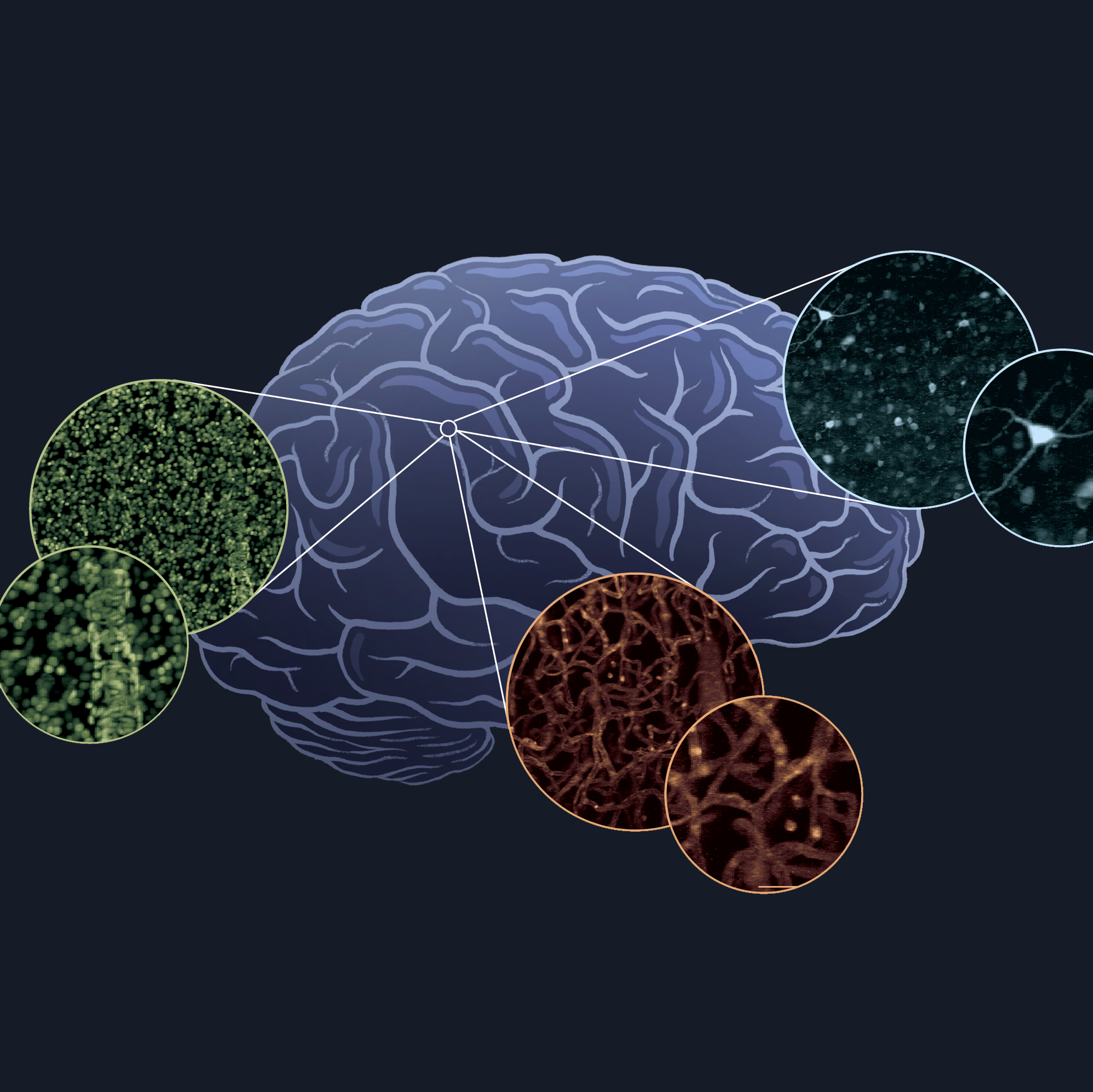
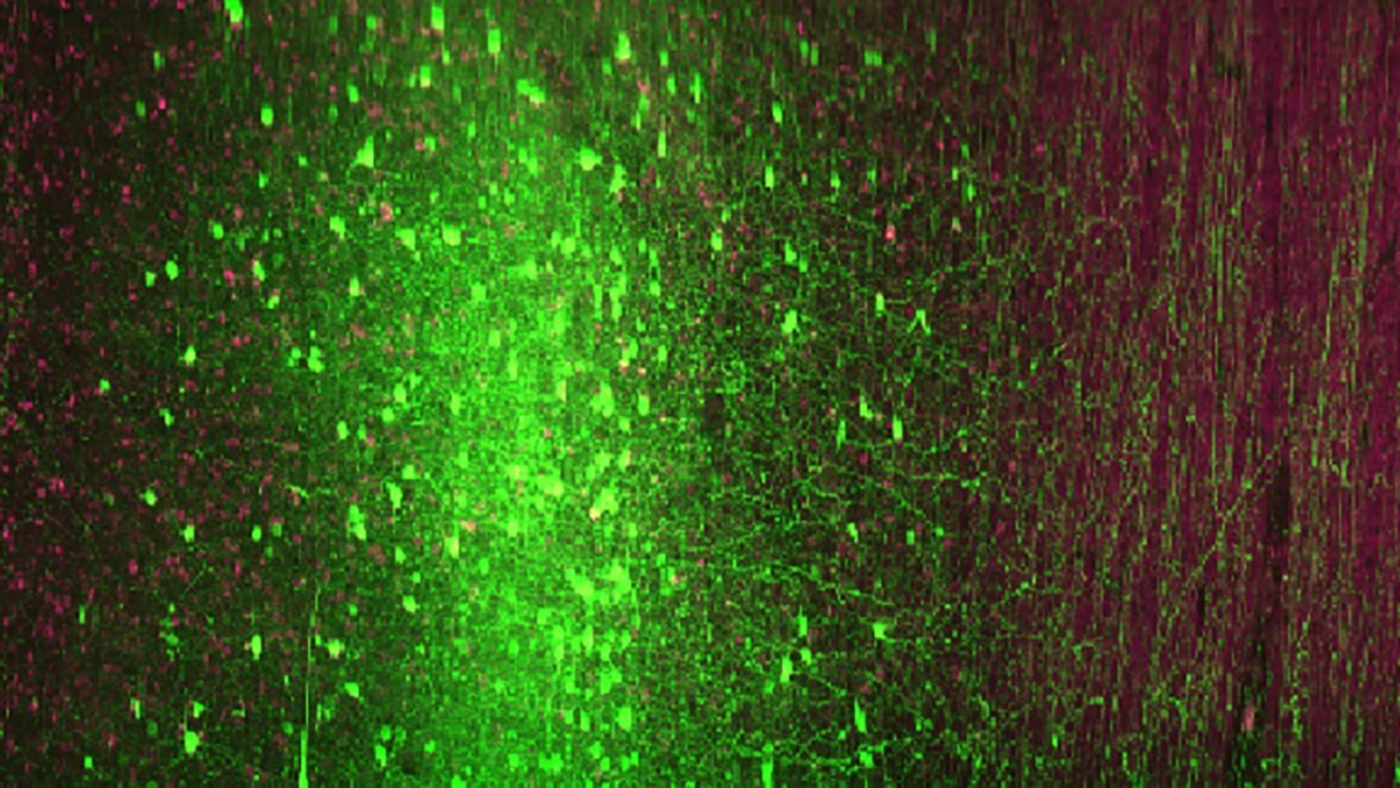
Advanced microscopy will help reveal the brain's wiring diagram. (Credit: The Hillman Lab)
“It’s extremely difficult to track connections between brain cells, so this is a big intellectual and conceptual challenge,” said Dr. Hillman. “The microscopy innovations that my lab will develop will enable high-resolution tracing of the pathways of axons projecting throughout large brain areas in humans and animals.” This part of the project will also involve Hui Wang, PhD, at Massachusetts General Hospital, an expert in polarization-sensitive optical coherence tomography (PS-OCT), and will continue Dr Hillman’s synergistic collaboration with Zhuhao Wu, PhD, of Weill Cornell Medicine, a pioneer in the processing and labeling of human brain tissue.
Combined with other powerful imaging techniques, such as advanced magnetic resonance imaging (MRI) and X-ray based Hierarchical Phase-Contrast Tomography (HiP-CT), the new LINC Center is expected to create new frameworks for analyzing, on massive scales, the cellular connectivity in individual brains. That’s important, Dr. Hillman said, both for understanding individual variability in human brains and for making medical treatments for brain disorders more effective.